Cochlear Implant
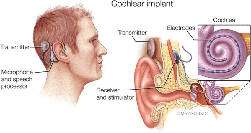
3min ReadA cochlear implant is a small electronic device that electrically stimulates the cochlear nerve (nerve for hearing). The implant has external and internal parts.
The external part sits behind the ear. It picks up sounds with a microphone. It then processes the sound and transmits it to the internal part of the implant.
The internal part is placed under the skin behind the ear during an outpatient surgery. A thin wire and small electrodes lead to the cochlea, which is part of the inner ear. The wire sends signals to the cochlear nerve, which sends sound information to the brain to produce a hearing sensation. Although normal hearing is not restored, with appropriate therapy and practice, the improved hearing experience can mean an increased awareness of sounds in the environment, as well as better communication through easier lip reading and listening.
Before surgery considerations
Primary care doctors usually refer patients to ear, nose and throat doctors (ENT doctors or otolaryngologists) to test them to see if they are candidates for cochlear implants.
Tests often done are:
• examination of external, middle, and inner ear for signs of infection or abnormality
• various tests of hearing, such as an audiogram
• a trial of hearing aid use to assess its potential benefit
• exams to evaluate middle and inner ear structures
• CT (computerized tomography) scan. This type of x-ray helps the doctor see if the cochlea has a normal shape. This scan is especially important if the patient has a history of meningitis because it helps see if there is new bone growth in the cochlea that could interfere with the insertion of the implant. This scan also may indicate which ear should be implanted.
• MRI (magnetic resonance imaging) scan
• psychological examination to see if the patient can cope with the implant
• physical exam to prepare for general anesthesia
During the Surgery
Cochlear implant surgery is done in a hospital or clinic. The surgery lasts two to four hours. You are given medication (general anesthesia) to make you sleep during the procedure.
• The surgeon makes a cut behind the ear and then opens the mastoid bone.
• The surgeon identifies the facial nerves and creates an opening between them to access the cochlea, which is then opened. He or she inserts the implant electrodes into the cochlea.
• The surgeon places an electronic device called the receiver under the skin behind the ear, securing it to the skull in this area.
• The incisions are then closed, and you will be moved into the recovery area and watched closely.
• You will be discharged after at least two hours of observation.
After the Surgery
Recovery duration after cochlear surgery is about 3 to 5 days, the patient might experience mild pain in ear, dizziness, or nausea which is the conclusion of anesthesia.
After a week, the stitches will be removed, and examinations must be done by the surgeon. About four to six weeks after the surgery, the external parts of the cochlear implant will be added.
Some important tips about the surgery
The patient will also learn the basics of using and caring for the implant. He may need to return for several visits over a few days for adjustments. Further fine-tuning will take place over several months. Learning to use a cochlear implant is a gradual process. It will likely require visits with speech-language pathologists and audiologists. With commitment, the patient can experience an improved quality of life with a cochlear implant.
Whom are qualified for this surgery?
Deep low hearing or deaf people are the most qualified ones.
Hospitalization duration
1 day hospitalization is necessary for this process


Reviews
Number of pending reviews175